

Biology Quiz: Name the 2 types of endoplasmic reticulum (ER).
Tell us what you think about our new Biology Quiz shorts, and subscribe to our new Biology channel to get more quizzes and biology animations!
#shorts #biology #cells
11
views
Biology Quiz: Which organelle transports proteins to the Golgi body?
Tell us what you think about our new Biology Quiz shorts, and subscribe to our new Biology channel to get more quizzes and biology animations!
#shorts #biology #cells
15
views
Scientific Method
For Employees of hospitals, schools, universities and libraries: download up to 8 FREE medical animations from Nucleus by signing up for a free trial at: http://nmal.nucleusmedicalmedia.com/biology_youtube
#ScientificMethod #ScienceFoundation #biology
SCIENCE ANIMATION TRANSCRIPT: Well begin our study of biology with the scientific method. The word biology is made up of the terms bio, which means life, and the suffix, ology, which means the study of. So, biology is the study of life and living organisms. And the foundation of all sciences, including biology, is the scientific method. The scientific method is an orderly way of investigating and evaluating factual information to learn how the world around us works. The basic steps of the scientific method are:making an observation, forming an inference and developing a hypothesis, conducting a controlled experiment, and drawing conclusions. Scientists use their senses to notice things in the world around them. After making observations, scientists ask questions, and then gather information, called data. After making observations, scientists ask questions, and then gather information, called data. For example, My car is terrible! is an example of subjective data. In contrast, My car wont start! is objective data, and is an example of an observation. Lets use this car trouble as an everyday example of using the scientific method to solve a problem. In this situation, when youre having trouble with your car, you might then ask, Why wont my car start? Well, there could be a number of causes maybe the car is out of fuel, or the battery might be dead. You could use an inference to possibly eliminate one of these things. Using an inference means to apply information in a logical way to reach a conclusion. In this example, your inference might be that the problem isnt a lack of fuel, because you know you filled the gas tank yesterday and havent driven the car very far since then. Your inference that a lack of fuel isnt the cause of the car failing to start may lead you to think the problem might be a dead battery. You can use this idea to form a testable explanation, called a hypothesis. You must to be able to test a hypothesis in order for it to be considered valid and scientific! A hypothesis can be presented in the form of an if then statement. In this case, the hypothesis might be, If my car starts when I use jumper cables, then the battery is the problem. This hypothesis is testable because either your car will or wont start when you use the jumper cables. Now, you can design a controlled experiment to test your hypothesis. During a controlled experiment, you control, or keep constant, all the factors, known as variables, except for the one you want to test. In this experiment, the variable that changes is the battery, while all other possible variables that might prevent the car from starting are not manipulated or changed. Why must you change only one variable? Because if you change or affect more than one, you wont know which variable caused the car to start. You can carry out your controlled experiment by attaching jumper cables from a charged battery to the battery in your car to see if the car then starts. In an actual experiment, you would record any data that results, such as how long you tried to jump-start the battery, and whether or not the car started. Well cover more details on controlled experiments and types of data in separate videos. After completing your experiment, you can draw a conclusion by using the resulting data to see if it supports your hypothesis. Remember, your hypothesis was, If my car starts when I use jumper cables, then the battery is the problem. The results of the experiment confirmed the hypothesis, so the conclusion is that the battery was the problem. If your car didnt start when using jumper cables, then your original hypothesis was not supported. As a result, a new hypothesis needs to be formed and tested. The scientific method continues until no more options remain. In review, the basic steps of the scientific method are: making an observation, forming an inference and developing a hypothesis, conducting a controlled experiment, and drawing conclusions.
NSV16035
29
views
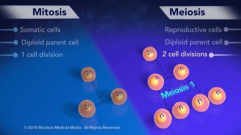
Mitosis vs Meiosis
For Employees of hospitals, schools, universities and libraries: download up to 8 FREE medical animations from Nucleus by signing up for a free trial at: http://nmal.nucleusmedicalmedia.com/biology_youtube
#mitosis #meiosis #CellDivision
SCIENCE ANIMATION TRANSCRIPT: Let's compare two types of cell division, mitosis and meiosis. While mitosis occurs all over the body in somatic cells, meiosis only occurs in the reproductive cells of the gonads in order to form gametes. The original cell in both mitosis and meiosis is diploid. Mitosis consists of one cell division, while meiosis consists of two stages of cell division called meiosis 1 and meiosis 2. Mitosis results in two diploid daughter cells. In contrast, meiosis results in four daughter cells that are haploid gametes. The two daughter cells resulting from mitosis are genetic duplicates of each other and the original cell. But each haploid gamete resulting from meiosis is genetically different from every gamete ever formed. [music]
NSV15013
14
views
Meiosis
For Employees of hospitals, schools, universities and libraries: download up to 8 FREE medical animations from Nucleus by signing up for a free trial at: http://nmal.nucleusmedicalmedia.com/biology_youtube
#meiosis #CellDivision #biology
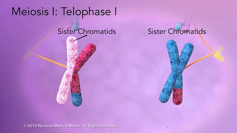
SCIENCE ANIMATION TRANSCRIPT: In this lesson, we'll explore the details of what happens during the phases of meiosis. Meiosis, sometimes called reduction division, is the type of cell division that produces gametes. By gametes, we mean sex cells such as sperm cells in males and egg cells in females. Meiosis is broken down into two stages of cell division called meiosis I and meiosis II. Meiosis I has four phases: prophase I, metaphase I, anaphase I, and telophase I. And meiosis II also has four phases: prophase II, metaphase II, anaphase II, and telophase II. Let's look at what happens during meiosis I. Prophase I starts with a diploid cell. Its chromatin contains two uncoiled, spread out sets of chromosomes, one from each parent. After the DNA in the chromatin replicates, it condenses into the more familiar X-shaped chromosomes. The replicated DNA is the same in the identical sister chromatids of each chromosome. In a process called synapsis, each chromosome pairs up with and binds to its corresponding homologous chromosome, forming a tetrad. A tetrad is the group of four sister chromatids in paired homologous chromosomes. The chromosomes contain genetic information called genes. These genes were inherited from each parent, and different versions of the same gene on each chromosome are called alleles. In a process called crossing over, chromatids from each homologous chromosome exchange segments of alleles. Also called recombination, crossing over randomly happens on every chromosome, resulting in different gene combinations. This explains why every gamete is genetically different from every other gamete. Crossing over results in genetic variety in offspring. This is why children are different from their biological parents, as well as from their biological siblings. Continuing on with prophase I, the nuclear membrane disappears, the centrioles move to opposite ends of the cell, and spindle fibers fan out from them. Next, in metaphase I, the homologous chromosomes line up at the equator and attach to spindle fibers from opposite poles. During anaphase I, spindle fibers separate the homologous chromosomes in each tetrad and pull them to opposite poles of the cell. The cell enters telophase I with one chromosome from each homologous pair at separate poles. However, each chromosome still consists of sister chromatids. Keep in mind that each chromosome's sister chromatids are no longer identical because of the allele exchange that happened during crossing over. Then spindle fibers disappear and the nuclear membrane re-forms around the chromosomes. Finally, cytokinesis occurs. Meiosis I ends with two genetically different haploid daughter cells. Each haploid cell contains only one set of chromosomes consisting of paired sister chromatids. Both cells now enter the next stage, meiosis II. However, unlike meiosis I, DNA does not replicate before meiosis II begins. Once again, in prophase II, the nuclear membrane disappears, and spindle fibers fan out from the two sets of paired centrioles. During metaphase II, the chromosomes in each cell line up at the equator and attach to spindle fibers from both poles. During anaphase II, the sister chromatids of each chromosome separate and move to opposite poles. Once the sister chromatids separate, they are called chromosomes. Finally, during telophase II, the spindle fibers disappear, and nuclear membranes re-form, and cytokinesis occurs in both cells. Meiosis II ends with four genetically different haploid daughter cells, each containing only one set of chromosomes. Some key points to remember about meiosis. It begins with a diploid cell. Meiosis only produces gametes. Gametes are genetically different haploid cells, sperm cells in males and eggs in females. Meiosis has two stages of cell division called meiosis I and meiosis II. During meiosis I, homologous chromosomes separate to produce two haploid cells, each containing chromosomes in the form of paired sister chromatids. In meiosis II, the sister chromatids separate in both cells, becoming individual chromosomes. Cytokinesis of these cells produces four genetically different haploid gametes. And here are some key points to remember about prophase I. The pairing of homologous chromosomes called synapsis occurs. Each pair of homologous chromosomes, consisting of four chromatids, is called a tetrad. During the process of crossing over, chromosomes in homologous pairs exchange segments of alleles. Crossing over results in genetic differences in gametes. All gametes produced by meiosis are haploid. [music]
NSV16019
25
views

Overview of Meiosis
For Employees of hospitals, schools, universities and libraries: download up to 8 FREE medical animations from Nucleus by signing up for a free trial at: http://nmal.nucleusmedicalmedia.com/biology_youtube
#meiosis #CellDivision #biology
SCIENCE ANIMATION TRANSCRIPT: In this lesson, we'll look at an overview of the type of cell division called meiosis. Meiosis takes place in an organism's reproductive structures called gonads for the sole purpose of producing haploid gametes that are genetically different. These haploid gametes are sperm cells in the male and eggs in the female. In the plant kingdom, pollen grains contain the male gamete, while structures called ovules contain the female gamete. Let's take a brief look at how meiosis produces gametes. A cell about to undergo meiosis will have already replicated its chromosomes during interphase of the cell cycle. This original cell is diploid, which means it has two sets of chromosomes, one from each parent, sometimes written as 2n. Through two stages of cell division, meiosis produces four genetically different haploid gametes, sometimes written as n. For this reason, meiosis is also called reduction division. It reduces the total chromosome number in half. So, when the haploid sperm cell and haploid egg cell unite to form a zygote during fertilization, the diploid number of chromosomes is restored in the resultant zygote. We'll examine the details of meiosis one and two in a separate video. [music]
NSV15016
8
views
Complicaciones de la presión arterial alta
Si deseas ver más imágenes médicas en 3D con precisión científica, suscríbase a nuestro canal: https://www.youtube.com/user/nucleushealthvideose
MEDICAL ANIMATION TRANSCRIPT: Si usted o alguien que conoce padecen de presión alterial alta, este vídeo le ayudará a comprender qué es y porqué es importante mantenerla bajo control. Con el tiempo, la presión arterial alta dañará las paredes de sus arterias. Este daño puede ocasionar condiciones de peligro de muerte, por ejemplo, una arteria puede debilitarse y formar un engrandecimiento llamado aneurisma. La pared puede romperse y sangrar en el tejido que la rodea. En otro ejemplo, el daño a la pared de una arteria puede ingresar determinadas sustancias en la sangre como colesterol, grasa y calcio. Estás pueden acumularse para formar lo que se denomina placa. A medida que la placa crece, se deduce el flujo sanguíneo en la arteria. Las células sanguíneas pueden pegarse a la placa y formar montículos sólidos llamados coágulos. Los coágulos de sangre reducen aún más o bloquean completamente el flujo sanguíneo. Si esto sucede en el cerebro puede provocar un ACV. Si esto sucede en el corazón puede provocar un ataque cardíaco. En otro ejemplo, el daño en las arterias en los riñones puede reducir su capacidad de funcionar correctamente. Esto puede provocar una enfermedad renal. Como último ejemplo, el daño a las arterias hace que el corazón lata con mayor fuerza la presión arterial aún más y puede ocasionar una insuficiencia cardíaca. Si tiene preguntas acerca de la presión arterial alta o cualquier medicamento que le hayan recetado, consulte con su médico. Es importante tomar los medicamentos siguiendo las instrucciones e informar acerca de cualquier efecto secundario que pueda tener.
ANH16172es
4
views
What are Haploid and Diploid Cells?
For Employees of hospitals, schools, universities and libraries: download up to 8 FREE medical animations from Nucleus by signing up for a free trial at: http://nmal.nucleusmedicalmedia.com/biology_youtube
#HaploidCells #DiploidCells #biology
SCIENCE ANIMATION TRANSCRIPT: In this video, we will discuss haploid versus diploid cells. Haploid and diploid are terms that describe the number of sets of chromosomes in a cell. Haploid means the cell has only one set of chromosomes. And diploid means the cell contains two sets of chromosomes. In your body, sex cells called Gametes have a haploid number of chromosomes represented by symbol n. In humans, every gamete has one set of 23 chromosomes, so the haploid, or n, number in humans is 23. This is important, since the union of gametes during fertilization creates a diploid cell called a zygote with two sets of chromosomes for a total of 46. At fertilization, the chromosomes from each parent match up to become the new pairs of chromosomes in a zygote. Each pair contains one chromosome from the father and a corresponding chromosome from the mother. These pairs are called homologous chromosomes. Homologous chromosomes are similar in shape and size along with the same types of genes in the same locations. A diploid zygote will go through cell division many times to produce all the cells in the body of a fully developed baby. All body cells except gametes are referred to as somatic cells. In humans, somatic cells are always diploid, written as 2n, which means they have 2 sets of 23 chromosomes for a total of 46 chromosomes. Other organisms have somatic cells with different diploid numbers of chromosomes. But the gametes in these organisms are haploid, meaning they always have half the diploid number of chromosomes. So, how does cell division affect the number of chromosomes in daughter cells? Well, somatic cells only reproduce by mitosis, a type of cell division that results in two genetically identical diploid daughter cells. In contrast, meiosis is a type of cell division that only produces gametes. In meiosis, a diploid cell undergoes two cell divisions to produce four genetically different haploid gametes. We'll cover the details of meiosis in another video. In summary, diploid cells have two complete sets of chromosomes. One set from each parent. Diploid cells have twice the number of chromosomes as haploid cells. The two sets consist of pairs of homologous chromosomes. The diploid chromosome number is written as 2n. All somatic cells, whether they're skin cells, muscle cells, or leaf cells in a plant are diploid. Diploid cells reproduce only by mitosis. And gametes are never diploid. In contrast, gamete cells, which are always haploid, have only one set of chromosomes, which is half the diploid number. Since there's only one set of chromosomes there are no homologous pairs. The haploid chromosome number is written as n. All gametes are haploid. And haploid gametes form from diploid cells through meiosis, never through mitosis. [music]
NSV15017
31
views
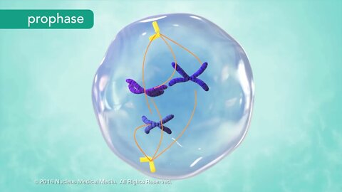
M Phase of the Cell Cycle
For Employees of hospitals, schools, universities and libraries: download up to 8 FREE medical animations from Nucleus by signing up for a free trial at: http://nmal.nucleusmedicalmedia.com/biology_youtube
SCIENCE ANIMATION TRANSCRIPT: In this lesson, we'll be exploring the M phase of the cell cycle including mitosis and cytokinesis. Let's do a quick review of the cell cycle to see where they fit in. The G1, S, and G2 phases make up interphase, and the M phase represents cell division. Cell division includes division of the nucleus, called mitosis, and division of the cytoplasm, called cytokinesis. Mitosis is further broken down into four phases: prophase, metaphase, anaphase, and telophase. Prophase is the longest phase of mitosis. Prophase is when chromatin begins to condense into the shape of chromosomes, and the nucleolus disappears. The previously replicated DNA coils tightly into sister chromatids. For the first time, you see individual chromosomes. In the center of each chromosome, a centromere attaches the sister chromatids together. Meanwhile, in the cytoplasm, microtubules known as spindle fibers begin to fan out from two sets of paired structures called centrioles. The spindle fibers elongate as the centrioles begin moving to opposite sides, or poles, of the cell. While this is happening, the nuclear membrane surrounding the nucleus disappears. Now that chromosomes are no longer separated from the cytoplasm, the opposite ends of the spindle fibers can attach to the centromeres. Next, the cell enters metaphase. The centrioles complete their movement to the poles of the cell while the spindle fibers line up the chromosomes along the equator of the cell. The end-to-end alignment of chromosomes results in a sister chromatid on either side of the equator. Anaphase follows metaphase. During anaphase, spindle fibers separate the sister chromatids at their centromere. Once separated from each other, each chromatid is called a chromosome. The single-stranded chromosomes form a V shape as the spindle fibers shorten and drag them through the gel-like cytoplasm. The chromosomes move to opposite poles of the cell toward their centrioles. It's common to confuse centrioles with centromeres which connect chromatids. Remember, centrioles are at the poles. Telophase is the final stage of mitosis. In telophase, a nuclear membrane re-forms around each set of chromosomes. Then the chromosomes spread out into chromatin, and the nucleolus becomes visible once again. Mitosis, the division of the nucleus, is now complete. The final step of the M phase is cytokinesis, the division of the cytoplasm. In animal cells, cytokinesis occurs through the inward movement of the cell membrane. This progressively pinches the cytoplasm until two identical daughter cells form. In contrast, plant cells can't pinch in two because they have a rigid cell wall surrounding their cell membrane. Instead, cell wall material assembles along the equator forming a structure called the cell plate. The cell plate grows until it joins with the existing cell membrane, separating the two halves of the cell into daughter cells. Over time, new cell walls form between the two daughter cells. Here are the key points to remember. The M phase is the fourth and final phase of the cell cycle. During the M phase, cell division occurs through two processes: mitosis, when the nucleus divides, and cytokinesis, when the cytoplasm divides. Mitosis has four phases. During prophase, chromatin condenses into chromosomes, spindle fibers form, and the nucleolus and nuclear membrane disappear. During metaphase, spindle fibers align the chromosomes along the cell equator. In anaphase, the spindle fibers separate sister chromatids into two separate groups of chromosomes, pulling them toward the poles. And in telophase, the nucleolus and nuclear membrane re-form. The chromosomes disperse into chromatin. Cytokinesis is division of the cytoplasm. The M phase is complete after cytokinesis occurs. The M phase of the cell cycle always results in two daughter cells. Both of these daughter cells are identical to each other and identical to the original cell that underwent mitosis. [music]
NSV15006
38
views
The Cell Cycle
For Employees of hospitals, schools, universities and libraries: download up to 8 FREE medical animations from Nucleus by signing up for a free trial at: http://nmal.nucleusmedicalmedia.com/biology_youtube
SCIENCE ANIMATION TRANSCRIPT: In this lesson, we'll be looking at the cell cycle. This is the lifespan of a eukaryotic somatic cell. A somatic cell is any cell in the body of an organism, except for sex cells such as sperm and egg cells. The cell cycle describes the sequence of cell growth and division. A cell spends most of its life a state called interphase. Interphase has three phases, the G1, S, and G2 phases. Interphase is followed by cell division, which has one phase, the M phase. Together these four phases make up the entire cell cycle. G1 of interphase is sometimes called growth 1 or gap phase 1. In G1, a cell is busy growing and carrying out whatever function it's supposed to do. Note that some cells, such as muscle and nerve cells, exit the cell cycle after G1 because they do not divide again. A cell enters the S phase after it grows to the point where it's no longer able to function well and needs to divide. The S stands for synthesis, which means to make, because a copy of DNA is being made during this phase. Once DNA replication is complete, the cell enters the shortest and the last part of interphase called G2, also known as growth 2 or gap phase 2. Right now, it's enough to know that further preparations for cell division take place in the G2 phase. Now that interphase is over, the cell is ready for cell division, which happens in the M phase. The M phase has two events. The main one is mitosis, which is division of the cell's nucleus, followed by cytokinesis, a division of the cytoplasm. So, at the end of M phase, you have two daughter cells identical to each other and identical to the original cell. Let's review. The cell cycle describes the life cycle of an individual cell. It has four phases, three in interphase and one for cell division. Most cell growth and function happen during G1. The cell enters the S phase when it needs to divide. In this phase the cell replicates its DNA. Replication just means the cell makes a copy of its DNA. In G2, the cell undergoes further preparations for cell division. Finally, we have cell division in the M phase. The M phase consists of mitosis, which is nuclear division, and cytokinesis, or division of the cytoplasm. We'll explore the details of mitosis and cytokinesis separately. [music]
NSV15004
38
views

Overview of Cell Division
For Employees of hospitals, schools, universities and libraries: download up to 8 FREE medical animations from Nucleus by signing up for a free trial at: http://nmal.nucleusmedicalmedia.com/biology_youtube
SCIENCE ANIMATION TRANSCRIPT: In this lesson, we'll be talking about how cells reproduce. How and why do they do this? Well, they use a process called cell division to make new cells called daughter cells. Unicellular organisms, meaning creatures that consist of just one cell such as bacteria, usually clone themselves during cell division. The two daughter cells that result are separate organisms, in this case, two new genetically identical bacteria. This is a type of asexual reproduction known as binary fission. Cells in a multicellular organism also reproduce by cell division, but the new daughter cells that are produced are not two separate organisms. Instead, these new cells are just parts of the organism, allowing it to grow, or sometimes replacing cells that are worn out or injured. For example, your body heals a paper cut through division of your skin cells, occurring at the edges of the cut. In a modified example of cell division, sex cells called gametes are made. Chromosomes are an important part of cell division. So, what are chromosomes? Let's look inside a cell's nucleus. Here we find the nuclear genetic material known as deoxyribonucleic acid or DNA. Each cell's DNA holds the genetic code or instructions from everything within that organism. Looking through a microscope, you can see that DNA is usually spread out within the nucleus. It looks kind of grainy. We call the DNA Chromatin when it looks like this. Before a cell divides, DNA must replicate or copy itself so that the information in this code can be passed on to each daughter cell. At the beginning of cell division, DNA condenses tightly into an x-shaped structure known as a chromosome. Each side of an x-shape chromosome is a genetically identical sister chromatid, forming a sideways v-shape. In the middle, a structure called a centromere, joins the sister chromatids together. Different types of organisms have different numbers of chromosomes. Every cell in your body is called a somatic cell, except your gametes. Human somatic cells have 23 pairs of chromosomes, for a total of 46. It's like the difference between how many pairs of shoes you have versus your total number of shoes. Gametes are the exceptions to this rule. Human eggs and sperm have only one chromosome from each pair, for a total of 23. And unlike somatic cells, gametes are not genetically identical to their parent cells. When a sperm cell fertilizes an egg, they both contribute their 23 chromosomes. This fused cell, called a zygote, now has 23 pairs of chromosomes for a total of 46. This overview of cell division will help prepare you for studying the cell cycle, which is the life cycle of the cell. [music]
NSV15003
50
views
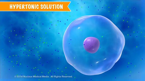
Cell Transport and Solutions
For Employees of hospitals, schools, universities and libraries: download up to 8 FREE medical animations from Nucleus by signing up for a free trial at: http://nmal.nucleusmedicalmedia.com/biology_youtube
#CellTransport #CellSolutions #biology
SCIENCE ANIMATION TRANSCRIPT: In this video, we'll discuss cell transport and solutions. The concentration gradient of extracellular solutions affects the transport of substances through the cell membrane. What can happen in this process? Well, cells may be surrounded by solutions with different particle concentrations, both in a lab as well as inside a living organism. So we need to know how cells will respond. Compared to the solution inside a cell, a solution outside the cell may be hypertonic, isotonic, or hypotonic. We'll describe hypertonic solutions using this beaker of water with dissolved salt, and a rather large cell submerged in it. To demonstrate water movement in and out of cells, we'll use simple numbers to illustrate concepts. Remember, the cell cytoplasm is mostly water as well. In this example, let's say the cytoplasm inside the cell is a solution containing 10% salt, which means the other 90% is water. Keep in mind percentages of substances within the cell must equal 100%. The percentages of solute and solvent in the beaker must also add up to 100%. However, the solution in the beaker is a different concentration from the solution inside the cell. It's 20% salt and 80% water. So you can see that there's a greater concentration of water inside the cell than outside, 90% compared to 80%. We call a solution hypertonic when its concentration of water is lower than inside the cell and its concentration of solute is higher than inside the cell. As a result, water molecules diffuse out of the cell through osmosis, causing the cell to shrivel a bit. Remember, osmosis means diffusion of water. If the solution's water concentration was significantly lower, the cell could shrivel to the point of imploding. For red blood cells in hypertonic solutions, the shriveling from osmotic water loss is called crenation. Let's look at an example of plant cells in a hypertonic solution. Normally, turgor pressure, which is the water pressure in a plant cell's central vacuole, helps support the cell wall and overall plant shape. When water leaves plant cells by osmosis, the cell membrane and its contents shrink away from the rigid cell wall, and turgor pressure decreases. This is called plasmolysis. Plasmolysis causes a plant to wilt. Let's look at another container, this time with a cell floating in an isotonic solution. Sometimes, the solution outside the cell has about the same concentration gradient as the concentration gradient inside the cell. Once again, in this example, the solution inside the cell is 90% water with 10% salt, but this time, the surrounding solution is also 90% water with 10% salt dissolved. We call a solution isotonic when its concentrations of water and solute are the same as inside the cell. But this doesn't mean that there is no movement. In an isotonic solution, the rate of water molecules entering the cell is equal to the rate of water molecules exiting the cell. The amount of water molecules going in equals the amount of water molecules going out. That's why you see the arrows pointing in two different directions. In an isotonic solution, the cell doesn't shrink or swell. It stays exactly the same size. A third possibility is a solution outside the cell with a higher concentration of water than the solution inside the cell. In this example, the solution inside the cell has 20% salt. So that means the remainder is 80% water, but the cell has been placed in a beaker that only has a 10% salt solution. Doing the math, we can see that the solution surrounding the cell is 90% water, which means there's a greater concentration of water outside the cell than inside. We call a solution hypotonic when its concentration of water is higher than inside the cell, and its concentration of solute is lower than inside the cell. So by osmosis, the water molecules will move passively into the cell until a state of equilibrium is reached. Hypotonic solutions cause a cell to swell up with water. If the water concentration outside the cell is high enough, the cell can swell to the point of bursting. This is called cytolysis. Cytolysis in red blood cells is called hemolysis. Cytolysis doesn't happen in plant cells because the rigid cell wall prevents the cells from bursting. Here's a little trick to remember that hypotonic solutions cause a cell to swell rather than shrivel. When you think of hypo, think of a big, swollen hippopotamus or hippo for short. So to recap, we can have solutions that are hypertonic, isotonic, or hypotonic with respect to the cell. In an isotonic solution, the water concentration inside and outside the cell stays about the same. The water concentration stays the same because the concentration of solute is the same...
NSV15018
110
views
Opciones de tratamiento para el cáncer de mama
Si deseas ver más imágenes médicas en 3D con precisión científica, suscríbase a nuestro canal: https://www.youtube.com/user/nucleushealthvideose
MEDICAL ANIMATION TRANSCRIPT: Usted o alguien que usted quiere puede haber sido recientemente diagnosticada con cáncer de mama, este video le ayudará a entender algunas de las opciones de tratamiento disponibles. El cáncer de mama es una enfermedad en la que algunas de las células de la mama empiezan a crecer sin control. Los tratamientos para el cáncer de mama pueden incluir: cirugía, radioterapia, quimioterapia, terapia hormonal, terapia dirigida e inmunoterapia. Hay varios tipos de cirugía según el tamaño y la ubicación del tumor. Este video no cubre todos los tipos de cirugía para cáncer de mama disponibles. Durante una tumorectomía se extirpa el tumor, junto con parte del tejido normal que lo rodea. En una mastectomía total o simple, se extirpa toda la mama. En una mastectomía radical modificada, se extirpa toda la mama, además se extirpan algunos de los ganglios linfáticos bajo el brazo y, a veces, otros tejidos del pecho también. Es posible que su médico también quiera administrarle radioterapia, esta terapia utiliza radiación para destruir las células cancerosas o impedir que crezcan. La radioterapia de haz externo utiliza una máquina que dirige la radiación hacia el cáncer desde fuera del cuerpo. La radioterapia de haz interno, también conocida como braquiterapia, utiliza una sustancia que emite radiación a través de diferentes tipos de dispositivos de administración, la sustancia se coloca dentro de su seno donde el cáncer se ha eliminado. La quimioterapia utiliza ciertos medicamentos para eliminar el cáncer, estos medicamentos pueden tratar las células cancerosas en todo el cuerpo o pueden administrarse para que traten las células cancerosas de un área. Otra opción de tratamiento es la terapia hormonal, las hormonas son sustancias que se encuentran naturalmente en el cuerpo; el estrógeno es una de estas hormonas. Su cuerpo utiliza el estrógeno principalmente para el crecimiento y desarrollo del útero, las mamas y los ovarios, sin embargo, el estrógeno también puede promover el crecimiento de ciertas células cancerosas que son receptoras hormonales positivas. Un tipo de terapia hormonal bloquea el estrógeno para que no se una a las células del cáncer de mama y así no haga que estas células crezcan y se dividan. La terapia dirigida es una opción de tratamiento para los cánceres que tienen un tipo de marcador específico en las células cancerosas; una terapia está dirigida a las células con marcadores llamados HER2. La HER2 es una proteína natural en las células que normalmente promueve el crecimiento saludable de las células. Sin embargo, algunas células cancerosas tienen demasiada cantidad de esta proteína, esto hace que las células cancerosas crezcan y se propaguen de forma más agresiva que las células normales. Los medicamentos de terapia dirigida están diseñados para unirse solo a las proteínas HER2 de las células cancerosas, como resultado las células cancerosas dejan de crecer y pueden morir. La inmuno oncología, también conocida como inmunoterapia, ayuda su sistema inmunitario a combatir el cáncer. El cáncer, a veces, se puede ocultar de las células inmunitarias que lo atacan, por ejemplo, tanto el cáncer como las células inmunitarias pueden tener proteínas, llamadas proteínas de punto de control, cuando estas se adhieren las otras células inmunitarias detienen su ataque. Los científicos buscan un tipo de medicamento de inmunoterapia que impida que las proteínas de punto de control se adhieran entre sí, como resultado, las células inmunitarias pueden atacar y destruir las células cancerosas. Su médico puede recetar una combinación de los tratamientos que hemos descrito u opciones de tratamiento diferentes a los mencionados aquí. Si tiene alguna pregunta sobre el cáncer de mama, o sobre cualquier medicamento que le hayan recetado consulte a su médico, es importante que tome sus medicamentos según las indicaciones y que informe cualquier efecto secundario que tenga.
ANH15164es
27
views
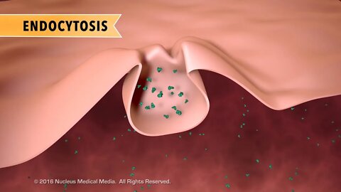
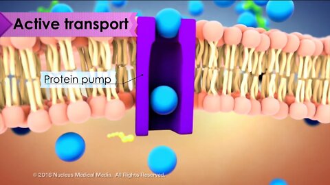
Cell Biology: Active Transport
For Employees of hospitals, schools, universities and libraries: download up to 8 FREE medical animations from Nucleus by signing up for a free trial at: http://nmal.nucleusmedicalmedia.com/biology_youtube
SCIENCE ANIMATION TRANSCRIPT: In this video, we'll discuss active transport. Active transport is when particles move from an area of low concentration to high concentration. This is also known as moving against the concentration gradient. The key thing to remember is that active transport requires energy. If passive transport is like a ball naturally rolling down a hill, active transport is the opposite. You can get the ball back up the hill, but you're going to have to expend some energy to do it. Cells require this type of substance movement in order to function properly. For example, heart muscle cells responsible for making your heart beat, move molecules or ions against their concentration gradient. So, what are some of the main types of active transport? We have endocytosis, exocytosis, and protein pumps. Sometimes a cell uses active transport to pull in large particles using its cell membrane. This is called endocytosis. One type of endocytosis is called phagocytosis. This often happens when the cell takes in some type of nutrient. In another type of endocytosis called pinocytosis, the cell takes in fluids by creating pockets in the cell membrane. The cell can ingest a large amount of fluid this way by pinching off these cell membrane pockets into the cytoplasm. The opposite of endocytosis is exocytosis. Exocytosis is when something needs to exit the cell. The cell can remove large molecules or wastes this way by fusing the membrane bound vesicles containing them with the cell membrane, forcing them out of the cell. A good way to remember that exocytosis is a way for things to leave the cell is that it shares the first two letters with exit. You can also remember that endocytosis is a way for things to move into the cell because each shares the first two letters with enter. Sometimes the cell uses special protein pumps to move small molecules or ions against the concentration gradient into or out of the cell. An example of this is the sodium potassium pump. In this process, the pump uses energy in the form of ATP molecules to move sodium ions out of the cell and then move potassium ions into the cell. Protein pumps used an active transport require energy because the molecules or ions are moving from an area of low concentration to high concentration. In summary, active transport is when the cell uses energy to move substances in or out of the cell against the concentration gradient via endocytosis, exocytosis, or protein pumps. [music]
NSV15009
9
views
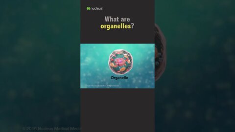
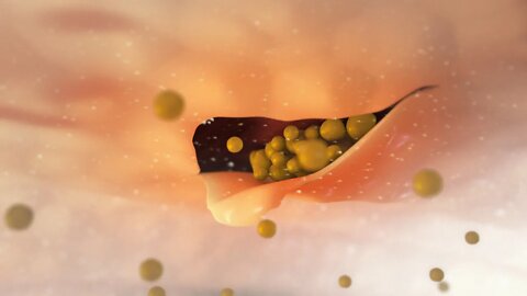
Biology Quiz: What are organelles?
Tell us what you think about our new Biology Quiz shorts, and subscribe to our new Biology channel to get more quizzes and biology animations!
#shorts #biology #cells
6
views
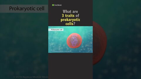
Biology Quiz: 3 traits of prokaryotic cells?
Tell us what you think about our new Biology Quiz shorts, and subscribe to our new Biology channel to get more quizzes and biology animations!
#biology #shorts #cells
4
views
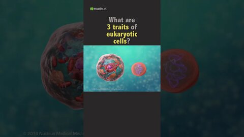
POP QUIZ: 3 traits of eukaryotic cells?
Tell us what you think about our new Biology Quiz shorts, and subscribe to the Nucleus Biology channel to get more quizzes and biology animations!
#shorts #biology #cells
1
view
¿Cómo se diagnostica la presión arterial alta?
Si deseas ver más imágenes médicas en 3D con precisión científica, suscríbase a nuestro canal: https://www.youtube.com/user/nucleushealthvideose
MEDICAL ANIMATION TRANSCRIPT: Un profesional médico puede medir su presión arterial para saber si tiene presión arterial alta, este video le ayudara a comprender qué significan los números en la presión arterial. Se puede medir la presión arterial con un dispositivo para la presión arterial denominado esfigmomanómetro. Cuando el corazón late, la presión de la sangre en las paredes de las arterias se denomina: presión sistólica; cuando su corazón se relaja entre latidos la presión en la pared arterial es llamada: presión diastólica. Su tensión arterial puede cambiar a lo largo del día, sin embargo, la tensión sistólica optima debería ser inferior a 120 y la tensión diastólica optima debería ser inferior a 80. Si su tensión sistólica con frecuencia se mantiene por encima de 130, o si su tensión diastólica con frecuencia se mantiene por encima de 80, usted sufre de tensión arterial alta. Sus objetivos específicos pueden variar dependiendo de la situación de su salud, pregúntele a su médico cuales deben ser los objetivos de su presión arterial.
ANH16170es
2
views

1 Minute Biology Quiz - 2 Categories of Cells
Subscribe to Nucleus Biology to prep for your next test! This #short is What are the 2 categories of cells?
#shorts #biology #quiz
1
view
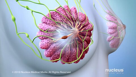
Comprender el Cáncer de Mama
Si deseas ver más imágenes médicas en 3D con precisión científica, suscríbase a nuestro canal: https://www.youtube.com/user/nucleushealthvideose
MEDICAL ANIMATION TRANSCRIPT: Usted o alguien que usted quiere puede haber sido recientemente diagnosticada con cáncer de mama. Este video le ayudará a entender qué es el cáncer de mama y cómo afecta a su cuerpo. Los senos son un par de órganos ubicados directamente debajo de la piel en el tórax. En el exterior de la mama está el pezón. El círculo de piel más oscura que lo rodea se llama areola. En las mujeres, los pechos están hechos de tejido graso, glándulas productoras de leche y tubos llamados conductos. Hay una amplia red de vasos linfáticos y ganglios linfáticos en la mama y alrededor de ella. El fluido de las mamas se drena a través de los vasos linfáticos hacia los ganglios linfáticos. Si el fluido contiene sustancias nocivas, como bacterias o virus, las células inmunes dentro de los ganglios linfáticos las atacarán y destruirán. Desde allí, la mayor parte del fluido pasa a los ganglios linfáticos que están debajo del brazo y luego a otros ganglios y vasos linfáticos que desembocan en el torrente sanguíneo. La mayoría de los cánceres de mama se inicia en los conductos de la mama, pero puede crecer en cualquier parte de la mama. Aquí se forman células cancerosas a partir de las células que recubren los conductos. Estas pueden crecer y multiplicarse para formar un tumor canceroso. Con el tiempo, las células cancerosas pueden propagarse a través de los ganglios linfáticos. Los siguientes pueden ser signos o síntomas de cáncer de mama. Tenga en cuenta que estos signos y síntomas no incluyen todos los casos. Durante un examen mamario de rutina, usted o su médico pueden sentir una pequeña protuberancia dura en su pecho o debajo del brazo. Además, es posible que salga un poco de líquido del pezón, o bien es posible que vea hoyuelos en la piel de la mama. Su médico usará ciertos términos para describir la evolución de su cáncer, llamada estadificación. La estadificación del cáncer de mama es compleja. Las siguientes descripciones de estadificación son una visión general y no incluyen todos los casos. La etapa 0 significa que se encuentran células anormales pero que no se han diseminado más allá de donde empezaron hacia otros tejidos de la mama. La etapa I significa un tumor menor de 2 centímetros en el tejido de mama. La etapa II A significa que la mama puede tener cáncer en los ganglios linfáticos de la axila, o bien puede ser un tumor de 2 centímetros o menos con cáncer en los ganglios linfáticos de la axila. La etapa II A también puede ser un tumor de entre 2 y 5 centímetros sin diseminación a los ganglios linfáticos. La etapa II B significa que la mama puede tener un tumor de entre 2 y 5 centímetros con cáncer en los ganglios linfáticos de la axila, o bien puede tratarse de un tumor mayor de 5 centímetros sin diseminación a los ganglios linfáticos. La etapa III A significa que la mama puede tener un tumor de cualquier tamaño y cáncer en los ganglios linfáticos de la axila, o bien puede ser un tumor mayor de 5 centímetros y cáncer en los ganglios linfáticos de la axila. La etapa III B significa que el tumor puede ser de cualquier tamaño y que el cáncer puede haberse diseminado a la pared del pecho, la piel de la mama y/o los ganglios linfáticos de la axila. La etapa III C significa que la mama puede no tener un tumor visible o un tumor de cualquier tamaño con diseminación a los ganglios linfáticos de la axila, el esternón o alrededor de la clavícula. La etapa IV significa que el cáncer se ha diseminado a órganos distantes. Usted puede preguntarse cómo ha llegado a tener cáncer de mama. Si bien es imposible predecir exactamente quién va a tener cáncer de mama, hay algunas cosas que pueden aumentar el riesgo: ser mujer, edad avanzada, terapia de reemplazo hormonal, tener el primer hijo después de los 30 años, exposición a la radiación en el pecho y un antecedente familiar de cáncer de mama. Esta lista no incluye todos los casos. Si se enfrenta a un diagnóstico de cáncer de mama, continúe conversando con su médico y su equipo de atención del cáncer.
ANH15163es
22
views
Análisis de colesterol en la sangre
Si deseas ver más imágenes médicas en 3D con precisión científica, suscríbase a nuestro canal: https://www.youtube.com/user/nucleushealthvideose
MEDICAL ANIMATION TRANSCRIPT: Si está viendo este video, su proveedor de atención médica le ha pedido a usted o a alguien que conoce que se someta a un análisis de sangre para medirse sus niveles de colesterol. Las pautas para el tratamiento del colesterol de los institutos nacionales de la salud recomiendan que se someta a un análisis de sangre cada 5 años si tiene 20 años o más. La prueba, llamada perfil lipoproteico en ayunas, mide sus niveles de colesterol total. LDL, también conocido como colesterol malo, HDL, también conocido como colesterol bueno y un tipo de grasa en su sangre llamada triglicéridos. Durante esta evaluación, se le tomará una muestra de sangre después de no haber comido durante 9 y 12 horas. Ayunar es importante para asegurar resultados precisos en la prueba. El objetivo del colesterol total debe ser menor a 200 miligramos por decilitro. Para la mayoría de personas, el HDL ideal, o colesterol bueno, debe ser de 60 o más. El LDL ideal, o colesterol malo, debe ser menor a 100. Y los triglicéridos en ayunas deben ser menores a 150. Si usted tiene niveles altos de colesterol en la sangre, tiene un riesgo mayor de desarrollar una enfermedad cardíaca. En la enfermedad cardíaca, los vasos sanguíneos, llamados arterias coronarias, se estrechan o bloquean con una sustancia serosa que contiene colesterol, llamada placa. Con el tiempo, la placa puede reducir o bloquear la corriente sanguínea rica en oxígeno que se dirige a su miocardio y causar un ataque cardíaco. Existen varios factores de riesgo para las enfermedades cardíacas: los factores de riesgo que usted puede controlar incluyen altos niveles de colesterol y triglicéridos en la sangre, presión arterial alta, diabetes, prediabetes, sobrepeso u obesidad, fumar, falta de actividad física, una dieta poco saludable y estrés. Los factores de riesgo que usted no puede controlar incluyen edad, género e historia familiar de enfermedades cardíacas. Si usted ya tiene una enfermedad cardíaca o factores de riesgo múltiples o graves, su médico puede reducir su objetivo de LDL a menos de 70. Su objetivo específico puede variar dependiendo en la situación de su salud. Pregúntele a su médico cuáles deben ser los objetivos de los lípidos.
ANH16169es
11
views
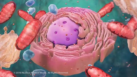
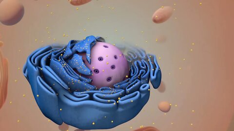
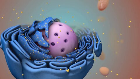
Overview of Cell Structure
For Employees of hospitals, schools, universities and libraries: download up to 8 FREE medical animations from Nucleus by signing up for a free trial at: http://nmal.nucleusmedicalmedia.com/biology_youtube
SCIENCE ANIMATION TRANSCRIPT: [music] Cells are the smallest living units of an organism. All cells have three things in common, no matter what type of cell they are. All cells have a cell membrane which separates the inside of the cell from its environment. Cytoplasm, which is a jelly-like fluid, and DNA, which is the cell's genetic material. There are two broad categories of cells. The first category is eukaryotic cells. They have organelles which include the nucleus and other special parts. Eukaryotic cells are more advanced complex cells such as those found in plants and animals. The second category is prokaryotic cells. They don't have a nucleus or membrane-enclosed organelles. They do have genetic material, but it's not contained within a nucleus. Prokaryotic cells are always one-celled or unicellular organisms, such as bacteria. [music] So, what are organelles? Organelle means little organ. Organelles are the specialized parts of a cell that have unique jobs to perform. Let's start with the nucleus, the control center of the cell. The nucleus contains DNA, or genetic material. DNA dictates what the cell is going to do and how it's going to do it. Chromatin is the tangled spread out form of DNA found inside the nuclear membrane. When a cell is ready to divide, DNA condenses into structures known as chromosomes. [music] The nucleus also contains a nucleolus, which is a structure where ribosomes are made. After ribosomes leave the nucleus, they will have the important job of synthesizing, or making, proteins. [music] Outside the nucleus, the ribosomes and the rest of the organelles float around in cytoplasm, which is the jelly-like substance. Ribosomes may wander freely within the cytoplasm or attach to the endoplasmic reticulum, sometimes abbreviated as ER. There are two types of ER. Rough ER has ribosomes attached to it. And smooth ER doesn't have ribosomes attached to it. The endoplasmic reticulum is a membrane-enclosed passageway for transporting materials such as the protein synthesized by ribosomes. Proteins and other materials emerge from the endoplasmic reticulum in small vesicles where the Golgi apparatus, sometimes called the Golgi body, receives them. As proteins move through the Golgi body, they are customized into forms that the cell can use. The Golgi body does this by folding the proteins into useable shapes or adding other materials onto them such as lipids or carbohydrates. Vacuoles are sack-like structures that store different materials. Here in this plant cell, the central vacuole stores water. [music] Going back to the animal cell, you will see an organelle called a lysosome. Lysosomes are the garbage collectors that take in damaged or worn out cell parts. They are filled with enzymes that break down the cellular debris. The mitochondrion is an organelle that is the powerhouse for both animal and plant cells. During a process called cellular respiration, the mitochondria make ATP molecules that provide the energy for all of the cells activities. Cells that need more energy have more mitochondria. [music] Meanwhile, the cell maintains its shape through a cytoskeleton. The cytoskeleton includes the thread-like microfilaments which are made of protein, and microtubules which are thin, hollow tubes. Some organisms such as plants that are photoautotrophic, meaning they capture sunlight for energy, have cells with an organelle called a chloroplast. The chloroplast is where photosynthesis happens. It's green because it has a green pigment called chlorophyll. Plant cells also have a cell wall outside of their cell membranes that shape, support, and protect the plant cell. Animal cells never have a cell wall. There are many other unique structures that only some cells have. Here are just a few. In humans, for example, the respiratory tract is lined with cells that have cilia. These are microscopic, hair-like projections that can move in waves. This feature helps trap inhaled particles in the air and expels them when you cough. Another unique feature in some cells is flagella. Some bacteria have flagella. A flagellum is like a little tail that can help a cell move or propel itself. The only human cell that has a flagellum is a sperm cell. In summary, remember, eukaryotic cells are plant and animal cells with a nucleus and membrane-enclosed organelles. While prokaryotic cells are unicellular organisms without these things. All cells have a cell membrane, cytoplasm, and genetic material. And even though only plant cells have chloroplast, both plant and animal cells have mitochondria. [music]
NSV15001
66
views
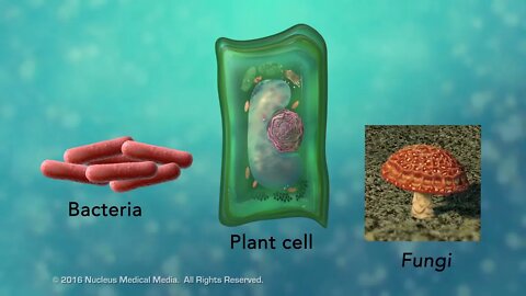
Overview of Cell Boundaries
For Employees of hospitals, schools, universities and libraries: download up to 8 FREE medical animations from Nucleus by signing up for a free trial at: http://nmal.nucleusmedicalmedia.com/biology_youtube
SCIENCE ANIMATION TRANSCRIPT: Today, we're going to talk about the outer boundary of cells. Every cell has a boundary to separate it from its surroundings. You may already know that plant cells have a rigid outer boundary called a cell wall. Other organisms such as bacteria and fungi also have cell walls. And while their cell walls differ in structure and composition, their cell walls all provide support, shape, and protection for these types of cells. It is essential you remember that animal cells always have a cell membrane but never have a cell wall. So what is the boundary that all cells have? Whether they have a cell wall or not, all cells have a cell membrane, also called a plasma membrane. In a typical animal cell, the cell membrane is a thin, flexible barrier against the outside environment. It's main job is to help with homeostasis, a type of equilibrium in which the cell maintains a relatively constant, stable internal environment. Like all living things, cells require stable internal conditions in order to survive, grow, and reproduce. The cell membrane helps maintain this stable internal environment by being selectively permeable. This means it acts as a gatekeeper to control or select what can get into or out of the cell. We'll learn more about the ways cells accomplish this separately. For now, remember that all cells have a flexible cell membrane, and most cells also have a rigid cell wall. And it's important to know that animal cells never have a cell wall. The cell wall provides support, shape, and protection to the cell. And the cell membrane is selectively permeable in order to help maintain intracellular homeostasis. [music]
NSV15002
12
views
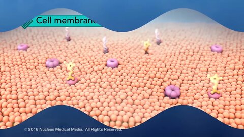
Structure of the Cell Membrane
For Employees of hospitals, schools, universities and libraries: download up to 8 FREE medical animations from Nucleus by signing up for a free trial at: http://nmal.nucleusmedicalmedia.com/biology_youtube
SCIENCE ANIMATION TRANSCRIPT: In this video, we will be discussing the structure of the cell membrane. When scientists looked at the selectively permeable cell membrane, they described its structure as a fluid mosaic. You might know that a mosaic is a picture made up of little tiles. Like a mosaic, the cell membrane is made up of different parts as well. The cell membrane has two layers of phospholipids referred to as a lipid bilayer. The lipid bilayer isn't rigid. The phospholipids in it have the ability to move in a flexible wave-like motion. Let's take a closer look at a few phospholipids. The round head portions are hydrophilic, which means they're attracted to water. Both the extracellular fluid, meaning fluid outside the cell, and the cytoplasm inside the cell are mostly made up of water. So, the hydrophilic phospholipid heads of the outer layer will be oriented toward the extracellular fluid. And the heads of the inner layer will be oriented toward the cytoplasm. The phospholipid tails are hydrophobic, which means watery areas withheld them. So they orient toward each other in a direction as far away from the watery content as possible. There are also scattered proteins embedded in the phospholipid layers, some with carbohydrates attached. So, in the fluid mosaic model, the cell membrane is made up of different parts. And these parts make up a flexible boundary around the cell. But how do the majority of substances get in our out of the cell? Some molecules sip through the little spaces in between the phospholipids, which make up the majority of the semi-permeable cell membrane. However, other molecules are too big to fit through the cell membrane this way. So, how do these larger molecules pass through the cell membrane? The molecules move through proteins embedded in the cell membrane, either from the extracellular area into the cell, or from the intracellular area out of the cell. These substances will move through tunnels made up of these proteins. We'll explore how things move through the cell membrane in greater detail separately. [music]
NSV15005
9
views
Overview of Cell Transport
For Employees of hospitals, schools, universities and libraries: download up to 8 FREE medical animations from Nucleus by signing up for a free trial at: http://nmal.nucleusmedicalmedia.com/biology_youtube
SCIENCE ANIMATION TRANSCRIPT: Cell transport is the process of how things move in or out of the cell through the cell membrane. There are two broad categories of cell transport. The first category is passive transport. For a cell, passive transport means it's an automatic process that doesn't require any input of energy. For example, diffusion is a passive process in which particles move either into or out of the cell from an area of higher concentration to an area of lower concentration. The cell doesn't use any energy when this happens. The second category of cell transport is active transport. This is when particles move from an area of lower concentration to an area of higher concentration. When particles move against the concentration gradient, energy is required often to allow protein pumps to assist in particle movement. Why would the cell need to move particles from a low to high concentration and expend energy to do it? An important example is seen in your heart muscle cells. In order for your heart to beat, there are certain molecules that have to move from an area of low concentration to an area of high concentration for those cardiac muscle cells to work. So, the main things to remember are passive transport happens automatically with no energy required, while active transport needs energy for it to occur. [music]
NSV15007
4
views