do you see what I see?
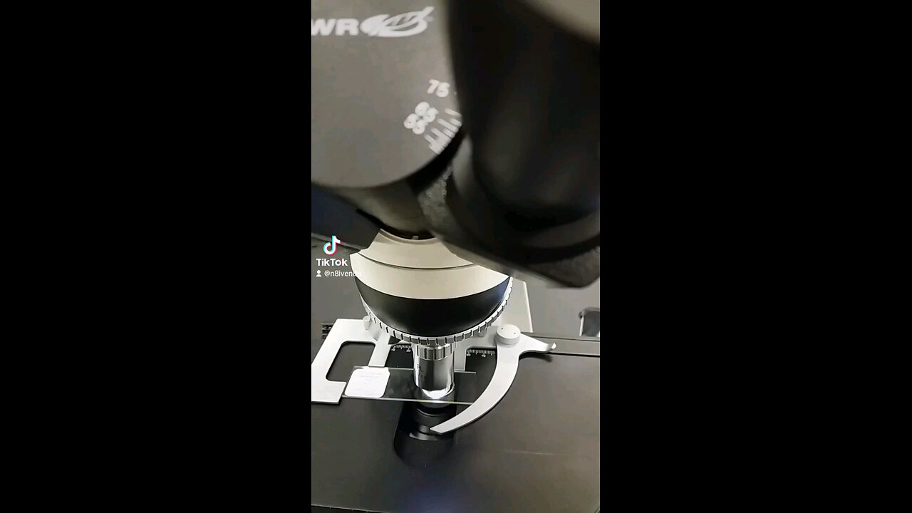
Under a microscope, a plant cell reveals a complex and highly organized structure, reflecting its role as the basic unit of plant structure and function. Let's explore the various components of a typical plant cell:
**Cell Wall:**
The first structure that catches the eye is the rigid cell wall, which provides structural support and protection to the cell. It's made up of cellulose, a complex carbohydrate. Under the microscope, the cell wall appears as a thick, outer layer surrounding the cell.
**Cell Membrane (Plasma Membrane):**
Inside the cell wall, there is a thin, flexible layer known as the cell membrane. It is composed of lipids and proteins and regulates the movement of materials in and out of the cell. The cell membrane is not always clearly visible under a light microscope but can be observed using more advanced microscopy techniques.
**Cytoplasm:**
The cytoplasm is a jelly-like substance that fills the cell and surrounds the organelles. It consists of water, enzymes, salts, and various organic molecules. Under the microscope, the cytoplasm appears as a granular material with small, moving particles known as cytoplasmic streaming.
**Nucleus:**
The nucleus is the control center of the cell and contains the cell's genetic material, DNA. It is surrounded by a double membrane called the nuclear envelope, which contains pores that regulate the passage of materials into and out of the nucleus. Inside the nucleus, DNA is organized into structures called chromosomes. Under the microscope, the nucleus appears as a dark-stained structure usually located near the center of the cell.
**Nucleolus:**
Inside the nucleus, there may be one or more nucleoli, which are dense, spherical structures composed of RNA and proteins. The nucleolus is involved in the production of ribosomes, the cell's protein-making machinery. Under the microscope, the nucleolus appears as a dark-stained region within the nucleus.
**Endoplasmic Reticulum (ER):**
The endoplasmic reticulum is a network of membrane-enclosed tubules and sacs that are involved in protein and lipid synthesis. There are two types of ER: rough endoplasmic reticulum (RER), which is studded with ribosomes, and smooth endoplasmic reticulum (SER), which lacks ribosomes. Under the microscope, the rough endoplasmic reticulum appears as a series of flattened sacs with small, dark-staining dots (ribosomes) attached to the surface.
**Ribosomes:**
Ribosomes are small, spherical organelles that are the site of protein synthesis. They can be found attached to the rough endoplasmic reticulum or floating freely in the cytoplasm. Under the microscope, ribosomes appear as small, dark-staining dots.
**Golgi Apparatus:**
The Golgi apparatus is a stack of flattened membrane-enclosed sacs that modifies, sorts, and packages proteins and lipids for transport within or outside the cell. Under the microscope, the Golgi apparatus appears as a series of flattened sacs arranged in a stack.
**Mitochondria:**
Mitochondria are membrane-bound organelles that are the site of cellular respiration, the process by which cells generate energy in the form of ATP. They have a double membrane, with an outer membrane and a highly folded inner membrane. Under the microscope, mitochondria appear as elongated structures with a smooth outer membrane and a highly folded inner membrane.
**Chloroplasts:**
Chloroplasts are membrane-bound organelles that are the site of photosynthesis, the process by which plants convert sunlight into energy. They contain chlorophyll, a green pigment that captures sunlight and uses it to produce sugars. Under the microscope, chloroplasts appear as green, oval-shaped structures containing stacks of membranous structures called thylakoids.
**Vacuole:**
The vacuole is a large, membrane-bound organelle that stores water, ions, and other molecules. It also helps maintain turgor pressure, which is important for maintaining the shape and rigidity of the cell. Under the microscope, the vacuole appears as a large, clear space surrounded by a membrane called the tonoplast.
**Plasmodesmata:**
Plasmodesmata are small channels that connect plant cells, allowing for the exchange of water, ions, and small molecules between adjacent cells. Under the microscope, plasmodesmata appear as small, dark-staining dots or lines that traverse the cell wall.
In summary, under a microscope, a plant cell reveals a complex and highly organized structure, including a cell wall, cell membrane, cytoplasm, nucleus, nucleolus, endoplasmic reticulum, ribosomes, Golgi apparatus, mitochondria, chloroplasts, vacuole, and plasmodesmata. Each of these structures plays a specific role in the growth, development, and functioning of the plant cell.
-
 21:37
21:37
Forrest Galante
13 hours ago6 Deadly Sea Monsters That Actually Exist
96.4K4 -
 LIVE
LIVE
JdaDelete
49 minutes agoElden Ring | First Playthrough Episode 10
75 watching -
 8:10
8:10
MattMorseTV
21 hours ago $55.65 earnedDemocrats caught COLLUDING with Epstein.
58.4K109 -
 LIVE
LIVE
Pepkilla
1 hour agoBreakfast First ~ Camo Grind Call Of Duty Black Ops 7
82 watching -
 LIVE
LIVE
DannyStreams
3 hours ago🟢 Live: Coffee & Tasking | 100 follower Grind
101 watching -
 2:03:42
The Connect: With Johnny Mitchell
23 hours ago $12.61 earnedAmerican Vigilante Reveals How He Went To WAR Against The WORST Cartels In Mexico
31.5K2 -
 LIVE
LIVE
Amish Zaku
2 hours agoArc Raiders
85 watching -
 17:04
17:04
T-SPLY
20 hours agoCongresswoman DENIED By Judge To Drop Federal Assault Charges!
29.2K37 -
 LIVE
LIVE
Astral Doge Plays!
1 hour agoFinal Fantasy X ~LIVE!~ The Dream Ends
52 watching -
 3:05:32
Game On!
23 hours ago $32.13 earnedCan We Reach 25,000 Followers? NFL Wiseguy Roundtable Week 11 Best Bets!
176K5