The Late Prof. Arne Burkhardt - Autopsies: Evidence for Jab Related Harm and Death. Feb. 5, 2022
The inaugural Understanding Vaccine Causation Conference, convened by World Council for Health- Prof. Arne Burkhardt full demonstration: https://rumble.com/v3yajuy-prof.-arne-burkhardt-autopsies-evidence-for-jab-related-harm-and-death-feb-.html?mref=1bxo9j&mrefc=20
The Late Prof. Arne Burkhardt joined the Medical Practice panel to share his presentation, Autopsies: Evidence for Jab Related Harm and Death. Feb. 5, 2022
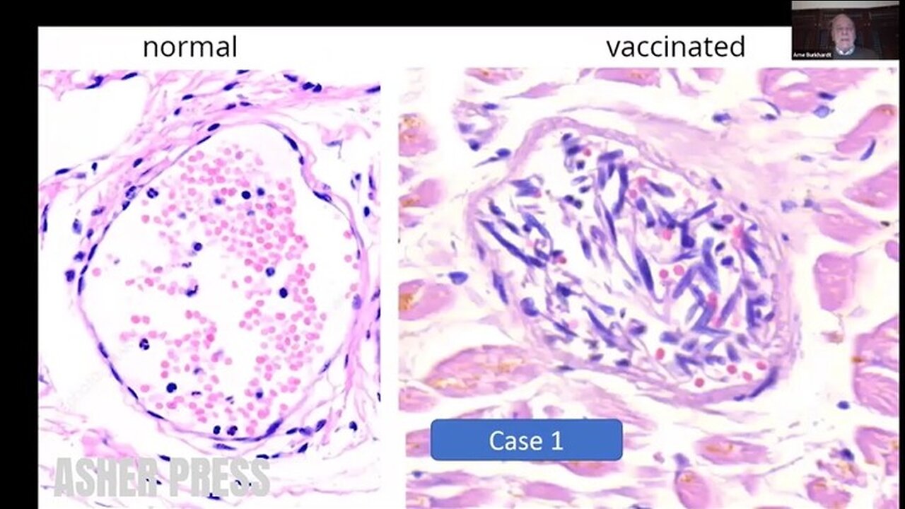
(Transcript) And I will just show you a few examples of the tissue damage that I have listed before. So here you can see a normal, small vessel, and you can see the Endothelium that is like a wallpaper and very small elongated spindle cell nuclei. And here in one of the cases, you can see that the Endothelium is in the lumen. And it is in there mixed with Lymphocytes and Erythrocytes and the nuclei is swollen.
So in some cases the small vessels even are completely destroyed by inflammatory infiltrates, mostly Lymphocytes. And this proves to me that it is an intravital reaction and not an autorhythmic phenomenon caused by degradation after death.
So we get the spike protein, immunohistochemistry on these cases and we see, you see here a very marked and specific, eh, the mark of the endothelium in these patients. And not only in the small vessels, but also in the smaller arteries, you can see it in the inner part of the vessel. And you can see here there’s decimated endothelial cells.
So not as I said, not only the smaller vessels were affected, but also the aorta and the larger arteries and two cases have died of a ruptured artery. And actually we found arteriosclerotic changes, but as you see here, it’s not very pronounced. But you can see inflammatory changes around in the deep layers of this aorta and also you can see some disturbance of structure of the smooth muscle and the elastic fibers. And if you have a higher magnification, you can see these small areas where the elastic fibers and smooth muscles are destroyed.
And again, lymphocytic infiltration proving that it was an intravital process. And here another case. We found it in, in five cases so this cannot be a coincidence. And we did the spike protein, and you find a marked positive expermeation of spike protein in the myofibroblasts of the arterial wall. This is the aorta. And also in the [unintelligible], you can see very strong expression of spike protein in these areas. And this I think is a very important finding.
So how often did we see this vasculitis, this endovasculitis or some call it endothelitis, in 11 cases with focal lymphocytic infiltration, then vasculitis paravasculitis in 10 cases, focal media-necrosis in six cases, and thrombosis caused on this area in two cases.
https://worldcouncilforhealth.org/multimedia/uvc-arne-burkhardt/
-
 1:50:51
Asher Press
1 day agoMike Benz - Epstein, CIA, Mossad, Trump - Triggernometry
413 -
 43:25
43:25
Michael Franzese
2 hours agoThis Is the Most Important Thing I’ll Ever Say
24.1K22 -
 LIVE
LIVE
Robert Gouveia
1 hour agoCongress HAMMERS Epstein Files! Maxwell APPEAL Decision! Obama Suspects LAWYER UP! 14th Amendment!
1,366 watching -

Barry Cunningham
3 hours agoPRESIDENT TRUMP TOURS JEROME POWELL SCENE OF THE CRIME AT THE FEDERAL RESERVE
30K9 -
 1:25:14
Sarah Westall
3 hours agoBlackmail, Power & Corruption: The Currency of the Empire - Epstein & more w/ Joachim Hagopian
20.4K5 -
 16:33
16:33
Clownfish TV
8 hours agoLate Night TV Has NO FUTURE After Stephen Colbert's Cancellation!
4K8 -
 45:56
45:56
The White House
3 hours agoPresident Trump Visits the Federal Reserve
31.6K19 -
 36:14
36:14
Kimberly Guilfoyle
4 hours agoRussia Hoax Reality Check, Live! | Ep240
29.4K10 -
 11:02
11:02
Preston Stewart
8 hours ago $0.61 earnedThailand–Cambodia Clash Erupts
10.5K3 -
 LIVE
LIVE
LFA TV
21 hours agoLFA TV ALL DAY STREAM - THURSDAY 7/24/25
948 watching