53. Craniocervical Junction Abnormalities (only) Dr. Harshfield
Resources from video chat:
12:42:22 From Doug Leonard : Craniosacral osteopaths know this and treat the junction all the time! Quess what I do
12:45:57 From Susan : what kind of adjustment was that?
12:46:32 From Russ Allen : Atlas Orthogonist
12:46:33 From Dr. Tom Lewis : C1 LOW VELOCITY ATLAS
Doug, is there an association which shows us how to locate local Craniosacral osteopaths?
12:51:04 From - : https://nucca.org/
12:51:09 From Dr. Tom Lewis : SWEAT INSTITUTE IN ATLANTA
12:51:23 From Louise Vogel : I am currently having my C1 repositioned by a chiropractor in PA, Marc Calicchio who trained w Dr. H’s colleague Dr. Scott Rosa. He doesn’t have the upright MRI and instead does x-rays which he interpolates from.
12:51:48 From - : http://www.sweatinstitute.com/t2/index.htm - Welcome To The Sweat Institute
12:51:52 From integrative brain : Are you utilizing Dr Rugierio's work on lymph drainage w/ Dr. D. Klinghardt. He has lymph cream and massage to open the pathway for brain drainage?
12:52:22 From Steve : Replying to "http://www.sweatinst..."
Thanx!
12:52:23 From - : https://nucca.org/ - Welcome to National Upper Cervical Chiropractic Association
12:52:58 From Doug Leonard : Replying to "Craniosacral osteopa..."
https://cranialacademy.org/
12:56:10 From - : What to Expect From Atlas Orthogonal Technique
- https://uppercervicalawareness.com/what-to-expect-from-atlas-orthogonal-technique/
13:01:23 From - : https://www.hudsonvalleyscoliosis.com/treatments/khan-kinetic-treatment/
13:03:00 From - : https://www.kktspine.com/kktstory/# - THE KKT TREATMENT PROCESS
13:03:16 From Dr. Tom Lewis : To get to Dr. Harshfield: [email protected]
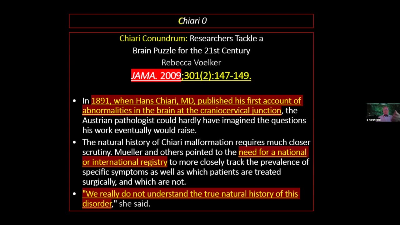
Craniocervical junction abnormalities are congenital or acquired abnormalities of the occipital bone, foramen magnum, or first two cervical vertebrae that decrease the space for the lower brain stem and cervical cord. These abnormalities can result in neck pain; syringomyelia; cerebellar, lower cranial nerve, and spinal cord deficits; and vertebrobasilar ischemia. Diagnosis involves magnetic resonance imaging (MRI) or computed tomography (CT).
-
 58:24
58:24
HEALTH EXPOSED By Health Revival Partners
4 months agoFoundational Healthcare to MAHA - Dr. Lewis
2342 -
 LIVE
LIVE
megimu32
1 hour agoOn The Subject: 90s Kid Sitcoms That Raised Us
107 watching -
 41:09
41:09
MattMorseTV
2 hours ago $32.14 earned🔴Top Dems. call for INSURRECTION.🔴
49.5K83 -
![Gray Zone Warfare [RGMT CONTENT Mgr. | RGMT GL | GZW CL]](https://1a-1791.com/video/fww1/02/s8/1/-/n/s/B/-nsBz.0kob-small-ARC-RAIDERS-FIRST-DROP-IN.jpg) LIVE
LIVE
XDDX_HiTower
48 minutes agoGray Zone Warfare [RGMT CONTENT Mgr. | RGMT GL | GZW CL]
57 watching -
 LIVE
LIVE
Charlotte Winslow
6 hours agoLet's Play DISPATCH!
51 watching -
 1:47:50
Side Scrollers Podcast
5 hours agoSide Scrollers Presents: OVERCOCKED
9.09K -
 LIVE
LIVE
tangodelta
1 hour agoGray Zone Warfare / Battlefield REDSEC - Fire For Effect
38 watching -
 LIVE
LIVE
Razeo
1 hour agoBald raider
25 watching -
 LIVE
LIVE
Adam Does Movies
8 hours agoBEST Stranger Things Recap Ever! - LIVE!
74 watching -
 13:10:29
LFA TV
1 day agoLIVE & BREAKING NEWS! | WEDNESDAY 11/19/25
191K26