Premium Only Content

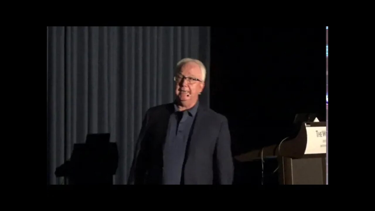
Feature Extraction in Bone Lesions Using CBCT Image Processing | Dale Miles, BA, DDS, MS
Feature Extraction in Bone Lesions Using CBCT Image Processing to Improve Diagnosis - from NICO to BRONJ | Dale Miles, BA, DDS, MS
IAOMT Annual Conference in Boston, MA
September 14, 2019
Learning Objectives:
1.Understand basic principles of CBCT.
2.View and understand which imaging filters and tools will help with diagnosis.
3.See these tools applied to bone lesions detected in the dental practice.
Dr. Miles has a full-time practice and is Adjunct Professor of Oral and Maxillofacial Radiology in the Department of Comprehensive Dentistry at the University of Texas Health Science Center at San Antonio. He was Chair of the Department of Oral Health Sciences at the University of Kentucky, the graduate program director of Diagnostic Sciences at Indiana University, and the first Associate Dean of Clinical Affairs at Arizona School of Dentistry & Oral Health. A diplomate of the American Board of Oral and Maxillofacial Radiology and the American Board of Oral Medicine, Dr. Miles has authored over 135 scientific articles, 6 radiology textbooks and the best-selling "Atlas of Cone Beam Imaging for Dental Applications."
Disclaimer: The information provided on this video is not intended as medical advice and
should not be interpreted as such. If you seek medical advice, please consult with a health care
professional. Also, the information in this video represents the thoughts of the individual
speaker/s, and the views expressed in this interview do not necessarily reflect the views of the
IAOMT, its individual members, its Executive Committee, its Scientific Advisory Council, its
administration, its employees, contractors, sponsors, or any other IAOMT affiliates.
-
 1:08:03
International Academy of Oral Medicine & Toxicology
2 years agoBoyd Haley, PhD, MIAOMT
346 -
 2:03:29
Tundra Tactical
8 hours ago $11.37 earned🛑LIVE NOW!! Honest Gun Company Slogans Gun Mad Libs and Much More
27.3K2 -
 4:54:33
MattMorseTV
9 hours ago $283.65 earned🔴Senate VOTES to END the SHUTDOWN.🔴
162K200 -
 2:55:39
Barry Cunningham
1 day agoBREAKING NEWS: DID PRESIDENT TRUMP MAKE A HUGE MISTAKE? SOME SUPPORTERS THINK SO!
59K40 -
 LIVE
LIVE
SpartakusLIVE
7 hours agoSOLOS on WZ || #1 Challenge MASTER is BACK in Verdansk
314 watching -
 2:49:38
megimu32
6 hours agoOFF THE SUBJECT: Chill Stream, Music & Fortnite Chaos 🎹🎮
39.5K4 -
 2:24:09
vivafrei
16 hours agoEp. 290: Canada's Darkest Week; Comey Fix is In! Tariffs, SNAP, Hush Money Win & MORE!
236K200 -
 5:01:48
EricJohnPizzaArtist
5 days agoAwesome Sauce PIZZA ART LIVE Ep. #68: DDayCobra Jeremy Prime!
33.8K12 -
 LIVE
LIVE
meleegames
6 hours ago32X Roulette - 30 Years. 32 Games. 32X.
81 watching -
 3:20:37
SOLTEKGG
5 hours ago(30+ KILL WORLD RECORD) - Battlefield 6
9.44K1